

Neurons are nerve cells that make up our nervous system. Unlike other cells, neurons are highly specialized to transmit information throughout our bodies.
We have 100 billion of these specialized nerve cells in our brain which is as many as the stars in the Milky Way. The ways to which they transmit the information, however, transcend the numbers of stars in the Universe.
Neurons vary in diameters from a range of 4 to 100 microns. They grow at a rate of 250,000 neurons/minute in utero but stop reproducing after birth. Know more about these nerve cells by getting to know its main types.
Types of Neurons
Interneurons
Interneurons consist of a large class of neurons that are found throughout the human body. Interneurons enable communication between sensory, or motor neurons, and the central nervous system.
They do this by creating neural circuits, which are groups of neurons that are interconnected by synapses and which carry out specific functions whenever they are activated. Interneurons function in reflexes, neuronal oscillations, and neurogenesis in adult brains of mammals.
Note: This post may contain affiliate links which will take you to online retailers that sell products and services. If you click on one and buy something, I may earn from qualifying purchases. See my Affiliate Disclosure for more details.
There are two types of interneurons:
- Local interneurons. Local interneurons form circuits with neurons close by and have short axons. They form these circuits in order to analyze small pieces of information.
- Relay interneurons. These neurons have long axons and they connect the circuits found in one region of the brain to circuits found in another region of the brain.
When you think of certain functions, including decision making and learning, they are only possible because of the interaction between these interneurons, which allows the brain to perform these complex functions.
Unlike the peripheral nervous system, the central nervous system – which includes the brain and spinal cord – contains a lot of interneurons. In the neocortex, which makes up roughly 80% of the human brain, 20-30% of the neurons are interneurons.
Following is a good way to think of interneurons. When you receive any type of physical shock, your body forms a reaction. The interneurons are responsible for this reaction, because they receive information from sensory neurons and send that data back to the motor neurons, which are the neurons that tell your body to respond to the shock.
In other words, interneurons are the link between motor neurons and sensory neurons, and they allow you to respond to stimuli or sensory input.
In other words, interneurons are middlemen neurons that are set between motor neurons and sensory neurons. The latter two neurons cannot communicate with one another without the interneurons sending messages back and forth between them.
Interneurons are located in the brain and spinal cord, but motor neurons and sensory neurons can be found all throughout your body.
If you get hit on the arm hard enough, it will hurt. In this instance, your sensory neurons send that feeling from the spinal cord to the brain, while the interneurons decide what to do about it.
They communicate their plan to the motor neurons, and they travel through your body to where the pain is and let you know about it so you can decide what to do next. If you decide to smack that person back, the motor neurons allow you to do this.
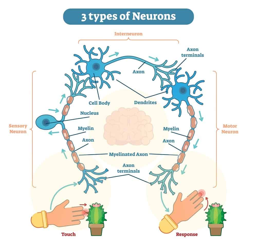
Neurons are also called nerve cells, and they are specialized cells whose main function is the transmission of various nerve impulses. Their cellular processes include dendrites and axons.
Dendrites are shorter processes located in a neuron’s cell body, and they are the processes that receive inputs from other neurons so that they can conduct the signals going to that cell body.
On the other hand, axons are longer and involve the singular process of relaying signals towards the tip – also called a synaptic terminal. In the brain of a human, there are over 100-billion interneurons, and one example of them is known as the Golgi cell, which is located in the cerebellum.
Integration is the official term that involves the process of the interneurons providing communication between the sensory and motor neurons so that some action can result.
Interneurons are like:
- … the local streets, which connect a neighborhood to a main road.
- … a club bouncer, because only the important “people” get by.
Interneurons are also known by the following names:
- Association neuron
- Connector neuron
- Intermediate neuron
- Internuncial neuron
- Local circuit neuron
- Relay neuron
Motor Neurons
Also called motoneurons, this is a neuron with a cell body that is located in the brainstem, motor cortex, or the spinal cord of the body. Its axon, or fiber, projects to either the spinal cord or outside of the spinal cord in order to directly or even indirectly control effector organs; in other words, glands and muscles.
Two types of motor neurons exist:
- lower motor neurons
- upper motor neurons.
The upper motor neurons have axons that synapse onto interneurons in the spinal cord. They occasionally synapse directly onto the lower motor neurons as well. The lower motor neurons are efferent nerve fibers, and their axons carry signals from the spinal cord to the effectors. There are also various types of lower motor neurons, including alpha motor neurons, beta motor neurons, and gamma motor neurons.
A single motor neuron can innervate many different muscle fibers, and any muscle fiber might undergo many action potentials in the short amount of time it takes for your muscle to twitch. When you think of the neurons doing their jobs, those jobs are completed in a very, very short period of time.
Muscles are usually stimulated repetitively so that the timing is just right, and for example, if that muscle twitch superimposes on another muscle twitch, several individual twitches can feel like one long twitch instead.
Motor columns of the spinal cord include:
- The median motor column, which runs the entire length of the spinal cord and which targets axial muscles.
- The hypaxial motor column, located in the thoracic region, which targets the body wall muscles.
- The preganglionic motor column, also located in the thoracic region, which targets the sympathetic ganglion.
- The lateral motor column, found in numerous places such as the lumbar and bronchial regions, which targets the muscles in the limbs.
- The phrenic motor column, located in the cervical region and which targets the diaphragm.
Upper and Lower Motor Neurons
The lower motor neurons start in the spinal cord and directly or indirectly innervate (supply an organ or body part with nerves) effector targets, meaning the glands and muscles. Their target varies somewhat, but the target is always some sort of muscle fiber in the somatic nervous system. There are three main categories of lower motor neurons, which are:
- General visceral motor neurons: these neurons indirectly innervate the smooth muscles of the viscera, or the muscles of the arteries, as well as the cardiac muscle. They are located within the peripheral nervous system, or PNS.
- Somatic motor neurons: originating in the central nervous system, these neurons project their axons to the skeletal muscles, that is, the muscles of the abdomen and limbs, which are involved in locomotion. There are three types of somatic motor neurons – the alpha efferent neurons, the beta efferent neurons, and the gamma efferent neurons. The word “efferent” is used to mean the flow of information to the periphery from the central nervous system.
- Special visceral motor neurons: also called branchial motor neurons, they are involved in actions that include phonation, swallowing, mastication, and even facial expressions. The associated cranial nerves include the abducens, trochlear, oculomotor, and hypoglossal nerves.
Upper motor neurons start in the motor cortex (part of the cerebral cortex located in the frontal lobe), which is located in the precentral gyrus. The cells in the primary motor cortex are Betz cells, which are giant neurons found in the gray matter of the brain, and they are a type of pyramidal cell.
In these cells, the axons descend from the cortex and form the corticospinal tract, which is located in the brain’s white matter and controls the limbs and the trunk.
Sensory Neurons
Sensory neurons are nerve cells that are located within the nervous system and which are responsible for converting external stimuli from the environment of the organism into electrical impulses that are internal.
This process is part of functions that include muscle contractions and even involuntary behaviors such as pain avoidance. These reflex circuits are usually found in the spinal cord in humans.
Also known as afferent neurons, sensory neurons convert a particular type of stimulus into action potentials or graded potentials via their receptors. The process is called sensory transduction.
The sensory neurons’ cell bodies are located in a part of the spinal cord known as the dorsal ganglia. Sensory information travels throughout the sensory nerve via afferent nerve fibers, which are nerve fibers that arrive at a particular region, not exit the region.
The information flows from the sensory nerve to the brain via the spinal cord. Stimuli can come from outside the body, including sound and light; or inside the body, including blood pressure or the sense of body position. Different sensory neuron types have different receptors that respond to different types of stimuli.
The different types and functions of sensory neurons include:
External Types of Sensory Neurons
- Taste: found in the taste receptors of the taste buds.
- Smell: olfactory receptor neurons that are activated by odor molecules found in the air.
- Vision: using photoreceptor cells that convert light into electrical signals, which are then refined and controlled with other types of neurons found in the retina. The five basic classes include bipolar cells, amacrine cells, photoreceptor cells, horizontal cells, and ganglion cells.
- Auditory: responsible for converting pressure waves that come from vibrating air molecules or sound into signals which can then be interpreted by the brain.
- Temperature: sensory receptors that respond to varying temperatures. Most experts agree that mammals have at least two specific types of thermoreceptors. These include the bulboid corpuscle, which detects cold temperatures; and warmth-sensitive receptors, which detect warm temperatures.
- Mechanoreceptors: these are sensory receptors that respond to mechanical forces, including distortion or pressure. There are several types of mechanoreceptors, including proprioceptors and nociceptors, with the latter which is responsible for processing temperature and pain changes.
Internal Types of Sensory Neurons
- Blood: these types of receptors are polymodal, meaning they respond to many different stimuli, and they can even detect changes in the chemical properties of the blood, including the concentration of oxygen.
- Nociceptors: these receptors send signals to the brain and spinal cord in response to stimuli that are potentially damaging. The receptors are found in both internal organs and on the surface of the body. There are three main types of nociceptors – chemical, which are involved in detecting some of the spices in certain foods; mechanical, which respond to both mechanical deformities and excessive pressure; and thermal, which are activated by noxious cold or heat at various temperatures.
The sensory system has some interesting quirks, including:
- Roughly 90% of a child’s knowledge comes from listening to conversations going on in the background, so even a slight amount of hearing loss can cause a child to fail at least one grade level.
- Approximately 80% of what you taste is directly affected by what you smell. Therefore, if you close your nose while eating a food you don’t like, you have a good chance of not tasting it as much.
- Every few days, your taste buds die off and regenerate. As you get older, this regeneration process slows down and can even result in the taste buds being dulled. This may be one reason why older people prefer salt and spices on their food.
- Every hour, the human eye can process over 35,000 pieces of information. In essence, your eyes transfer data to the brain for processing so that it can be instantly accessed.
- Humans have more receptors for pain than they do anything else. Pain is important because it is your body’s warning system, which is one of the reasons why masking pain before you exercise is usually not a good idea.
- The most sensitive parts of the body include the fingertips, soles of the feet, lips, and the back of the neck. One of the least sensitive parts is the middle of the back, even though this is the area that many massage therapists concentrate on the most.
Types of Nervous Systems

Central Nervous System (CNS)
Made up of the brain and spinal cord, the CNS is all nerve that is encased in bone.
Peripheral Nervous System (PNS)
Consisting of all of the other nerves in your body, including all of the nerves which are not encased in bone, the PNS consists of two main categories: the somatic nervous system and the autonomic nervous system.
- The autonomic nervous system controls the body’s functions which are automatic. Think of functions such as lungs, heart rate, and your internal organs, if you eat some junk food, you don’t have to think about squirting stomach acid on that food item or turning the food into fat and glucose that your body uses for other functions. The autonomic nervous system does this for you. In other words, any function in your body that is automatic and not something you yourself control is part of the autonomic nervous system. In addition, the autonomic nervous system is divided even further into:
Parasympathetic nervous system
This is a part of the autonomic nervous system that relaxes us. If you eat an unusually large supper, you are tired, the blood hangs out in your stomach, and your pupils constrict, so life is good at least for the moment. This is the parasympathetic nervous system at work.
Sympathetic nervous system
This nervous system does the opposite of the parasympathetic system. If you get nervous, upset, or even anxious, the sympathetic nervous system goes into action. It speeds up your heart rate, moves a lot of your blood into your arms and legs and away from your stomach, and dilates your pupils.
The body thinks that when you are in a state of stress, there is a possibility that you could die, so it gets ready to either fight or runs. This is known as the fight or flight response, and it is what the sympathetic nervous system does best.
- The somatic nervous system controls all muscle movements that are voluntary, including scratching an itch, kicking a ball, and reaching for the TV remote. Therefore, when you choose to make your muscles move a certain way, you are using the somatic nervous system’s motor neurons to accomplish the task.
Fun Facts about the Nervous System

- The central nervous system, or CNS, is comprised of the brain, spinal cord, and the retina of the eyes. The skull protects the brain and the skeletal vertebrae protect the spinal cord.
- The peripheral nervous system, or PNS, consists of all of the other nervous system structures located outside of the CNS, but which help connect the CNS to various areas in the body.
- The nervous system is very complex and is, in essence, the electrical wiring of the human body.
- All humans and animals that have backbones and spinal columns have a nervous system of vertebrates that are divided into two main parts – the CNS and the PNS.
- Your nervous system can transmit its signals at speeds of over 300 feet per second.
- In addition to the 100-billion neurons in the human brain, there are also 13.5 million neurons in the spinal cord of a human being.
- Sensory neurons change touch, sound, and light signals into neural signals, which are then sent back to the CNS in order to help the body react to and understand its surroundings.
- Motor neurons transmit their neural signals in order to activate glands or muscles.
- The nerves in your body are vulnerable to all types of damage, including damage through various diseases and even physical damage. If you damage a nerve, there is a lot of pain, as well as the loss of muscle control and loss of feeling in that particular area.
- Neurologists and neurosurgeons treat conditions of the nervous system, while physiatrists are the ones who help patients who have damage to their nervous system.
You might like our article about the “7 Different Types of Active Transport”
Glossary of Terms

Autonomic Nervous System: A part of the peripheral nervous system, it supplies neural connections to the glands and the smooth muscles of internal organs. Divided into the sympathetic and parasympathetic nervous systems, some experts consider the enteric system to be the third division.
Axon: The extension from a cell that carries nerve impulses from the body of the cell to other neurons.
Central Nervous System (CNS): The nervous system made up of the brain and the spinal cord.
Cerebellum: Located in the back of the brain, it controls basic movement, balance, and even posture.
Dendrite: One extension of a cell body which is made up of a neuron’s reception surfaces.
Gyrus: The raised portion of the convoluted surface of the brain.
Hypothalamus: Primarily involved in functions related to emotion, hunger, thirst, and control of the pituitary gland.
Limbic System: Controls primarily memory and emotions.
Motor Cortex: This is a section of the cerebral cortex, and it sends impulses to motor neurons. It is also involved in the coordination of movement and is located in the frontal lobe of the brain.
Myelin: The fatty insulation found surrounding the axon, it improves the speed of conduction of the nerve’s impulses.
Nervous System: A system that extends throughout the entire body and which connects every organ to the brain. Usually divided into the central nervous system (CNS) and the peripheral nervous system (PNS).
Neurotransmitter: A neurotransmitter is a chemical substance that is released at synapses which transmits information to the next neuron.
Parasympathetic Nervous System: One of two systems that make up the autonomic nervous system. Slows your heartbeat, constricts your airways, stimulates digestion, and constricts pupils, as well as other activities, for people in a relaxed state.
Peripheral Nervous System (PNS): This is a portion of the nervous system and includes all of the neurons and nerves located outside of the brain and spinal cord (i.e., outside of the central nervous system).
Primary Motor Cortex: This is the area that initiates voluntary movement.
Sympathetic Nervous System: One of two systems that make up the autonomic nervous system. It responds to stress or fear and causes dilated pupils, relaxed airways, accelerated heartbeats, and inhibited salivation, among others.
Synapse: The synapse is the area between one neuron and the next one. Neurotransmitters pass through them and transmit neural messages.

